Photoacoustic Imaging for Improved Surgical Guidance
A concern in surgeries like intranasal transhenoidal surgery is that when the surgeon is drilling through the sphenoid bone, they may accidentally rupture the carotid artery (which is just behind this bone). If surgeons were able to better determine where exactly the artery was behind the bone as they were drilling, then they could significantly reduce the risk of damaging the artery during surgery. Photoacoustic imaging can help in this scenario, because the acoustic response generated by an artery behind bone is different than when there is not an artery present behind the bone.
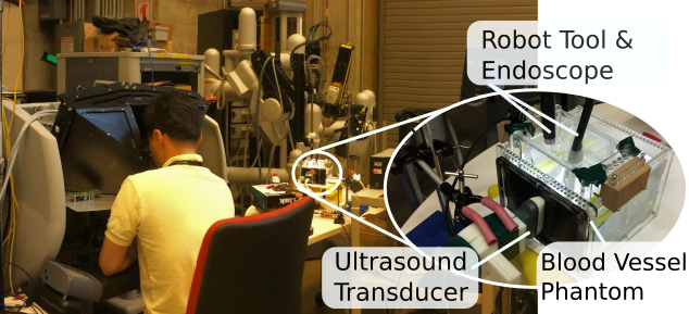
I worked on developing an experimental platform that could be used to show how effective such a technique could be. I designed and built a surgical phantom to simulate the carotid arteries behind the sphenoid bone, then analyzed the collected data to show how photoacoustic images could clearly show the presence of carotid arteries during surgery.

Publications#
- Photoacoustic‑Based Approach to Surgical Guidance Performed with and without a da Vinci Robot N. Gandhi, M. Allard, Sungmin Kim, P. Kazanzides, M.A.L. Bell. Journal of Biomedical Optics, 2017.
- Accuracy of a Novel Photoacoustic‑Based Approach to Surgical Guidance Performed with and without a da Vinci Robot N. Gandhi, S. Kim, P. Kazanzides, M.A.L. Bell SPIE BiOS, 2017.
- Improving the Safety of Telerobotic Drilling of the Skull Base via Photoacoustic Sensing of the Carotid Arteries S. Kim, N. Gandhi, M.A.L. Bell, P. Kazanzides ICRA, 2017.